Project Grant 2016
Multifunctional fiber optics
Principal investigator:
Fredrik Laurell, Professor of Laser Physics
Co-investigators:
KTH Royal Institute of Technology
Aman Russom
Michael Fokine
Ulf Österberg
Walter Margulis
Karolinska Institutet
Matthias Löhr
NTNU - Norwegian University of Science and Technology
Ursula Gibson
Institution:
KTH Royal Institute of Technology
Grant in SEK:
SEK 31.8 million over five years
Thank to fiber optics we can surf the internet and send huge quantities of data at high speed back and forth across the globe. Fiber optic cables use particles of light – photons – to transfer information, explains Fredrik Laurell, who is Professor of Laser Physics at KTH Royal Institute of Technology. Normally he works at AlbaNova University Center, but now he is guesting SciLifeLab in Solna, north of Stockholm, to meet some colleagues who are developing medical applications.
“Fiber optics is the backbone of modern-day communication. You might say that the twentieth century was the age of electronics, and that the twenty-first century is the era of photonics. Instead of electricity and electrons, new technology makes use of light and photons. The number of applications is constantly growing.”
One use of optical fibers and lasers is the study of cells and bacteria in vivo, i.e. in their natural environment. Laurell is heading a five-year project on multifunctional fibers funded by the Knut and Alice Wallenberg Foundation. The project involves collaboration with a number of researchers with different specialties in the field of optical physics.
“I like the multidisciplinary approach, and working with many different partners. The funding means we will have greater freedom in our research, and we can put together a larger research team. The Foundation’s support is also a testament to the quality of the work we are doing.”
Filling cavities with functions
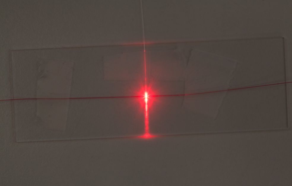
At the heart of fiber optics lie thin fibers made of silica. They are no more than 0.1 millimeters in diameter; inside they may contain various cavities and channels. These cavities can then be filled with semiconductors, fluids or metals to create different functions for use in biomedicine or electronics.
Laurell has worked for many years with researchers at RISE Acreo, formerly known as the Institute for Optical Research. His most important partner there is Walter Margulis, who is a guest professor at KTH, and is also taking part in the project on multifunctional fibers.
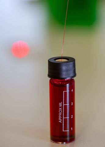
“As part of the project Acreo is making fibers with cavities of various kinds that we can fill and weld together in different combinations. Examples include electrodes to modulate the light, or channeling fluid into cavities resembling a classic capillary,” Laurell explains, pointing to an image on his computer illustrating how it might look.
“We can use light channeled to the center of the fiber to study the fluid. The amazing thing about this is that it’s all done on such a minute scale, but that it still works.”
Studying cells
Among other things, the project team wants to study the possibility of inserting the thin fiber into the body to examine cells and organs. Cancer treatments where fiber optics first find the cancer cells, and then deliver chemotherapy might be a potential application. Laurell Elaborates:
“This is the reason we’re collaborating with SciLifeLab and Professor Aman Russom, whose field is chip-based technologies for cell analysis and cell treatment. The project involves both basic and applied components. But clinical studies are still some time away.”
Cancer spreads via circulation of fluids in the body, particularly via blood and the lymphatic system. Finding these cells is like finding a needle in a haystack. The vision is to use fiber optic capillaries to distinguish diseased cells from healthy ones. When the technology has been further developed, the fiber optics will be tested by Professor Mattias Löhr at Stockholm’s Karolinska Institutet, who is researching on cancer of the pancreas.
“The pancreas is located behind the stomach, and is difficult to reach. But if we can get our fiber optics into it and feed cytostatic agents down the fiber channels, it would make diagnosis and treatment easier,” Laurell explains.
Removing cells
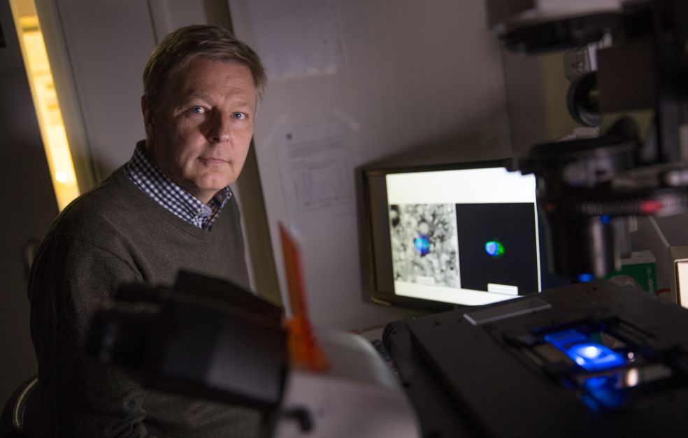
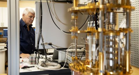
In the laboratory at SciLifeLab Laurell demonstrates a mini-flow cytometer, which uses fluorescence and fiber optics to identify and count specific particles in a stream of fluid. Traditional flow cytometers are much larger. This one is a simplified and miniaturized version of the technology.
“We built this one using fibers and mini-lasers instead. It has roughly the same capacity as the full-size version, but costs fifty thousand kronor instead of more than a million.”
The system can also be used to sort or remove particles from the fluid – cells as well as bacteria.
“So far we’ve only worked on cells that we have labeled using various fluorescent agents. We also want to develop a technique that works with autofluorescence – the light emitted naturally by cells, and other spectroscopic diagnostic methods.”
The fluid in the fiber can also be used as a laser medium, and if the cells are pumped into the fiber, there is potential to create a living laser in the fiber. Another exciting line of research being pursued by the team is how long-wave THz waves can be generated in fiber. This technique could be used for medical imaging inside the body, for example.
“It’s in the pipeline, and we’ve taken the first tentative steps,” Laurell adds.
Text Susanne Rosén
Translation Maxwell Arding
Photo Magnus Bergström
