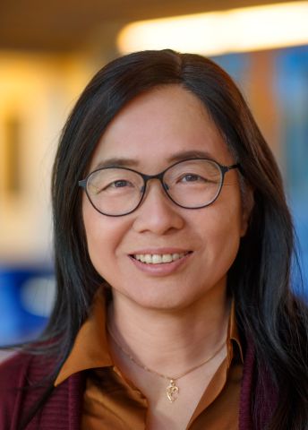
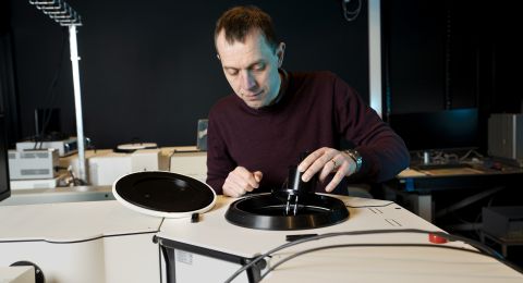
Xiaodong Zou has long dreamed of being able to take pictures of the tiniest constituents of nature, such as aromatic substances – scents and odors. She is now developing analytical methods that image small molecules in three dimensions and in greater detail than ever before. This opens the way for better drugs and new fundamental discoveries in chemistry.
Xiaodong Zou
Professor of Structural Chemistry
Wallenberg Scholar
Institution:
Stockholm University
Research field:
Structural chemistry and electron crystallography. Electron crystallography methods for mapping atomic structure in a material or a molecule.
As long ago as the 1990s, when she was a young PhD student, Zou wanted to image the structure of proteins, the smallest building blocks of life. But the technology at the time was not yet ready, and she had to shelve her plans.
“My supervisor said the project was too difficult, but I never let go of the idea.”
For many years she focused on inorganic materials, especially porous materials, and worked painstakingly on developing methods. Meanwhile, technological advances were being made. With the help of electron microscopy she could begin to obtain high-resolution 3D images of materials.
This opened a window to a new “nano-world” in which it is possible to see how the atoms that make up a material are arranged. This new knowledge has enabled scientists to tailor materials possessing the desired properties.
She continued to nurture the dream of being able to image organic molecules with the same richness of detail, one aim being to learn how different biological systems work and how to develop more effective drugs to treat diseases.
“The method available is called X-ray diffraction, but it has a number of drawbacks,” Zou explains.
To perform analyses using X-ray diffraction, the researchers have to reconstitute the substance into relatively large crystals, which is difficult with complex materials and biological molecules, which may contain thousands of atoms. X-ray diffraction usually requires access to a synchrotron. This makes the process expensive.
Fantastic images
But technological developments in recent years have caught up with Zou’s vision. She is now using electron diffraction to obtain increasingly detailed high-resolution images of crystals. The technique is quicker and more accessible; it can be installed “in-house,” and does not require such large crystals.
“Electron diffraction gives us fantastic images of crystals that are less than one micrometer across. By way of comparison, crystals tens of micrometers across are needed for X-ray diffraction. The reason is that electrons interact much more strongly with the atoms in the crystal than X-rays do,” Zou explains.
Much of her research involves methods development. Zou has researched a method called MicroED, which is used to study protein microcrystals.
One advantage of MicroED is that it can also provide information about atomic charge. This creates a completely new understanding of biochemical processes, since the proteins involved in the reactions often adopt different charge states as their atoms accept or donate electrons.
Crystalline sponges
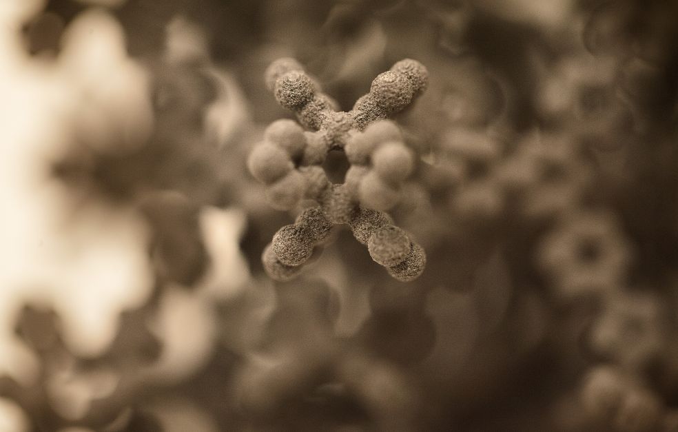
Zou is also working on a new method called Multi-CS, which may give a boost to the whole research field. Molecules are captured in a porous crystalline matrix – a “crystalline sponge,” enabling the researchers to analyze substances that were previously impossible to study. The original method was developed in Japan by Makoto Fujita and his research team in 2013.
“It’s a method that enables us to examine substances present in extremely small quantities or that are difficult to crystallize, such as oil and volatile substances,” says Zou.
The funding from Knut and Alice Wallenberg Foundation has been crucial to my career in Sweden. Being chosen as a Wallenberg Scholar means I can focus my research on key questions in the field of electron crystallography.
These may be new drugs, volatile aromatic compounds or even traces of hazardous substances.
The method has been used in combination with X-ray diffraction, but now Zou wants to develop the method by integrating it with electron diffraction. She is keen to develop a kind of screening strategy to create a library of crystalline sponges that are suitable for different molecules. New software and improved data analysis are also key components of the research.
“When we start to gather data from thousands of crystals it’s important to gain better control of data processing so we can get answers quickly.”
The Multi-CS method is extraordinarily sensitive, and the hope for the future is to analyze substances all the way down to picogram level – a trillionth of a gram.
Forensic medicine and customs
So far the research is purely scientific, but there are potential future applications. It might be possible to identify previously unknown substances, analyze metabolites in the body or trace pollutants in the environment with a precision that was previously inconceivable. Forensic medicine and customs are other areas where there is a need to trace substances, e.g. when analyzing unknown substances from a crime scene.
“And it might be possible to build a machine that captures molecules which could be used in airports,” Zou reflects.
The only boundaries are those set by the imagination. Meanwhile, Zou continues to work on the whole palette of methods. She is eager to optimize a broad range of tools, from microscopy and software to the collection and analysis of data. She hopes the technology will be a standard tool in many laboratories within a few years.
“We have seen a great interest among researchers and companies, and want to do our utmost to exchange ideas, educate students and build this field internationally.”
Text Nils Johan Tjärnlund
Translation Maxwell Arding
Photo Magnus Bergström
