Project Grant 2013
Karolinska CardioVascular Initiative
Principal Investigator:
Kenneth Chien, professor of cardiology
Co-investigators:
Karl-Henrik Grinnemo
Eva Hellström-Lindberg
Urban Lendahl
Institution:
Karolinska Institutet
Grant in SEK:
SEK 24.6 million over three years
Kenneth Chien has devoted all of his adult life trying to understand more about the heart's functions. He has worked at several leading biomedical institutions in the United States, most recently at Harvard, but in 2013 he moved his research group to Karolinska Institutet, KI.
“Sweden is an attractive research country, and KI has a special position. In many ways, KI is an undiscovered diamond with great future potential. I see no reason why Swedish research should not be able to compete with the world's finest top tiers and I think that more of my colleagues will find their way here over the coming years,” he says.
The benefits of doing research in Sweden are numerous, according to Chien. Here exists a relatively homogeneous population, interesting patient registries with high-quality data and more liberal regulations concerning embryonic stem cells compared to the US. At KI there is proximity between basic research and clinical studies, so-called translational research, and collaboration with an advanced pharmaceutical industry.
“In addition, all speak English, so you do not need to learn Swedish,” he says and laughs.
A blueprint of the heart
Now his own field, cardiovascular research, face a whole new era. One can begin to map and understand the molecular mechanisms of the heart. Chien's research aims to develop construction drawings, the blueprint for the heart. For a long time we know of the external structure, but now it's the inner layers you wish to access.
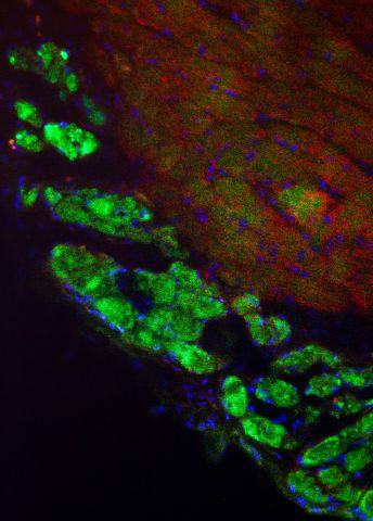
Chien and his colleagues have discovered that there are three types of stem cells in the fetus that gives rise to all cell types in different parts of the heart, such as the chambers of the heart, blood vessel walls and connective tissue. Now they want to in detail find out how the signal pathways look and how the cell types communicate to find the right part of the heart.
“Such detailed drawing gives us information about how the heart is constructed. It also means opening the door to repair a damaged heart. It's like when you renovate a house. One must consult the drawings for the renovation to be successful.”
Large differences between mice and humans
Previously one has been dependent on animal models. But there are large differences between, say, mice and humans. A mouse heart is created in just 48 hours, while the human heart is built up over several months. The human heart is ten thousand times larger than the mouse and significantly more dynamic and complex.
“It's like comparing a bike and a sports car,” says Kenneth Chien.” And so we must now begin to study the sports car, that is, move on to start studying humans as a model system.”
In Danderyd there are records of thousands of patients with hereditary cardiovascular diseases. It is a unique database and a gold mine for research. One can use the genetic information of these unusual heart diseases to understand how cells in the heart are coupled with each other. In the long run, it also provides opportunities to develop new treatments, advanced gene therapy for rare heart diseases.
Another research track is to use stem cell technology. In the near future Chien hope that it will be possible to cultivate cardiac muscle cells, cardiomyocytes, from human embryonic stem cells. Heart muscle cells can be purified with antibodies and then grow into functional tissue in animal models. At the horizon is the possibility that stem cell therapy can be tested in human trials.
“So far, the challenge has been to control the stem cells to become just the kind of heart cells that we wished, in large quantities and organized into functioning muscle units in the intact living heart,” says Kenneth Chien.

Existing cells can repair heart
A third form of treatment may in the future be to encourage existing cells in the heart to repair the damaged parts. Normally scars occur while the heart is healing after an injury. By providing the right chemical signals in a technology called synthetic m-RNA one could induce heart stem cells to proliferate in the intact heart and form new muscle tissue. In other words, the heart heals itself by expressing the protein in the heart muscle directly.
“We have managed to repair the hearts of mice by expressing a single protein that directs the heart stem cells away from being scar and towards becoming functional heart and vascular tissue. The injection made the treated mice survive two to three times longer than those who had no treatment. Now we have to find out if the corresponding technique can work in humans.”
The first human studies of this synthetic mRNA is slated for late in 2015, and the work is done stepwise. First attempts are made on skin before moving on to more vital organs such as the heart.
Kenneth Chien is enthusiastic about the new technological opportunities. What just a few years ago were considered science fiction is soon to be part of ordinary research.
“It's a dream come true as we can seriously begin to understand the heart and explore future treatments. But without the financial support to take on high risk-high reward science, we would go nowhere. Here the Knut and Alice Wallenberg Foundation plays a hugely important role as research funding provider, not the least through the strategic decisions to invest in daring research and to provide for the next generation, the young promising researchers.”
Text Nils Johan Tjärnlund
Translation Karolinska Institutet
Photo Magnus Bergström
